CTOS 2023
Connective Tissue Oncolcogy Society (CTOS) Conference 2023 (Dublin) Update Notes by Maisie England
This year’s annual CTOS conference was held in Dublin and was well attended by many medical professionals and patient advocates from across the world.
During the 4 day event, we met with many to talk through their specific LMS research and it gave a strong sense of hope for leiomyosarcoma due to the the continued breakthroughs in science. LMSDR will continue to work with researchers across the world to help further research which will directly benefit our community.
We had more detailed conversations with researchers on areas such as: Hot Tumour microimmune environments to help refine immune biomarkers and support patients in selecting the best clinical trials for them; treatment resistant Polyaneuploid cancer cells (PACCs) and how we can seek to identify the best way to target and weaken such cell groups; the SUNRISE LMS trial and what is next for the study; and candidate markers and molecular pathways associated with response and resistance to Gem/Tax therapy in LMS.

Over the years, the LMSDR foundation has supported many researchers on LMS specific work https://www.lmsdr.org/grant-awards and the CTOS 2023 conference offered an opportunity for us to meet with researchers such as Dr Matt van de Rijn, Dr Joanna Przbyl and Dr Paul Huang on their research. We discussed what is next for Liquid Biopsy and biological pathways research.
We also met with Dr Sant Chawla to discuss recent clinical trials such as the TT1-621 phase I/ II trial where TTI-621 acts by binding human CD47 and preventing it from delivering an inhibitory "do not eat" (anti phagocytic) signal to macrophages (see also https://clinicaltrials.gov/study/NCT02663518). Though this trial is still currently underway, there are positive results, especially where there are benefits due to low toxicity.
With Dr Chawla, we also discussed the Phase 2 study: A metronomic regimen using gemcitabine, doxorubicin, docetaxel and nivolumab for advanced leiomyosarcoma (NCT04535713) which concluded that: Taken together, the data suggests that nivolumab plus metronomic doses of gemcitabine, doxorubicin, and docetaxel (1) may have synergistic activity, and (2) is a promising second/third line combination regimen for advanced leiomyosarcoma with manageable toxicity. (See Poster 117). Overall, it was noted about the positive response rate and the ability to keep on this treatment for a longer period of time.
We had further conversations with Dr Jonathan Fletcher on the Strategic Advances in Sarcoma Science (SASS) network and its conference from 2023. The network is a North America and European network in which LMSDR is one of the sponsors. Dr Fletcher noted the key outcomes/ deliverables from the main sessions. In particular, the support for multi-disciplinary opportunities for early career researchers/ young investigators and how they have built in further support into the network to encourage more collaborative working.
He highlighted that they are currently setting the agenda for the next conference in 2024 and will potentially be focusing on LMS as one of the conference breakout topics.
In summary, the CTOS 2023 conference was a fantastic opportunity for LMSDR to understand what science research continues and what is in the future horizon for exciting new research we can support which continues to work towards a cure of LMS.
The Annexes note some of the specific LMS trials that are being ran and further LMS research that is being explored (as highlighted at the CTOS 2023 conference).
ANNEX 1:
RESEARCH POSTERS WITH SPECIFIC RELEVANCE TO LMS
(P104) COMPREHENSIVE IMMUNE PROFILING UNVEILS A SUBSET OF LEIOMYOSARCOMA WITH HOT TUMOR IMMUNE MICROENVIRONMENT
Objective: To investigate the immune biomarker in Leiomyosarcoma (LMS) which is a rare and recognized as an “immune-cold” cancer showing a poor response rate (< 10%) to immune checkpoint inhibitors (ICIs). However, durable response and clinical benefit to ICIs observed in few LMS, including, but not only, LMS with tertiary lymphoid structure (TLS) structures.
Methods: We used comprehensive transcriptomic profiling and a deconvolution method extracted from RNA-sequencing gene expression data in two independent LMS cohorts, the International Cancer Genome Consortium (ICGC, N=146) and The Cancer Genome Atlas (TCGA, N=75) to explore tumor immune microenvironment (TIME) in LMS.
Results: Unsupervised clustering analysis using the previously validated two methods-- 90 gene signature and the Cell-type Identification By Estimating Relative Subsets of RNA Transcripts (CIBERSORT), identified immune hot (I-H) and immune high (I-Hi) LMS respectively in ICGC cohort. Similarly, immune active groups (T-H, T-Hi) were identified in TCGA cohort using these two methods. These immune active (“hot”) clusters were significantly associated, but not completely overlapping, with several validated immune signatures such as sarcoma immune class (SIC) classification and TLS score, T cell inflamed signature (TIS) score, immune infiltration score (IIS), and macrophage score (M1/M2), with more patients identified by our clustering as potentially immune hot.
Conclusion: Comprehensive immune profiling revealed a subset of LMS with a distinct active ("hot”) TIME, consistently associated with several validated immune signatures in other cancers. This suggests that the methodologies that we used in this study warrant further validation and development, which can potentially help refine our current immune biomarkers to select the right LMS patients for ICIs in clinical trials.

(P 113) IMMUNOSARC II: A SPANISH SARCOMA GROUP (GEIS) PHASE IB TRIAL OF DOXORUBICIN AND DACARBAZINE PLUS NIVOLUMAB IN FIRST LINE OF ADVANCED LEIOMYOSARCOMA
Objective: The immunogenic cell death (ICD) caused by certain chemotherapy agents consists of molecular changes in tumor dying cells that stimulate immunogenicity and enhance antitumor effects. Doxorubicin is a drug with recognized ICD induction capacity, mainly through calreticulin membrane translocation that elicits immunogenic signals as phagocytosis by dendritic cells. The combination of doxorubicin and pembrolizumab was explored in a phase I/II trial treating patients with metastatic/unresectable sarcomas, achieving Partial Response (PR) in the 22% of patients, stable disease (SD) in the 59%, and progressive disease (PD) in the 19%, with median progression-free survival (mPFS) of 8.1 months, superior to historical controls. We hypothesized that the addition of an anti-PD1 (nivolumab) would increase the antitumor activity of doxorubicin plus dacarbazine, a commonly used upfront polychemotherapy regimen in advanced leiomyosarcoma (LMS) . We present here the phase Ib, cohort 7b of IMMUNOSARC trial.
Methods: Adult patients, with good performance status (ECOG 0-1), naïve of previous anthracycline-containing treatments and with centrally confirmed diagnosis of advanced/metastatic LMS were eligible. Initial dose level 0 (L0) was defined as doxorubicin (DOX) 75 mg/m2/day in 20 min on D1 followed by dacarbazine (DAC) 400 mg/m2/day in 60 min on D1 and 2, plus nivolumab (NIV) 360 mg on D2 after DAC every 21 days with GCSF support. This combo would be given up to 6 courses of 21-day cycles, followed by 1-year NIV maintenance. A -1 dose level (L-1) was defined with the same regimen but with NIV 240 mg. A classic 3+3 phase 1 design was used to determine the recommended phase 2 dose (RP2D) based on dose-limiting toxicities (DLTs), which was the main endpoint, observed during the first 21-day cycle. An expansion of the cohort with RP2D up to 20 evaluable patients was foreseen. Secondary endpoints included overall response rate (ORR), safety profile, translational objectives, among others. A baseline tumoral biopsy and blood sampling baseline and every 6 weeks were mandatory for translational purposes. Levels of expression of HMGB1 were assessed in plasma at week 6 and compared with baseline levels.
Results: Between January 2022 and February 2023, 20 patients (6 Male/14 Female), ECOG 0/1 (15/5), with median age 54 years (range 31-72) were enrolled. All patients were treated with the initial L0 scheme and no DLTs were observed being L0 the RP2D. Grade 3-4 toxicities were neutropenia (20%), anemia (10%), febrile neutropenia, asthenia, alopecia, and GGT increased (5% each). Four patients were not evaluable for efficacy at the moment of the data lock (1 due to uncompliant dosing and in 3 patients the first tumor assessment was not reached yet at the data lock). Of 16 efficacy-evaluable patients, RECIST ORR according to local clinical site assessment was 9 PR (56.3%), 6 SD (37.5%), and 1 PD (6.3%) (2 SD cases showed tumor size reductions >20%). Five patients ended treatment due to progression (4 radiological, 1 clinical) and 15 patients remain under the trial therapy. With a median follow-up of 8 months (2-12), in the 16 evaluable patients, the mPFS was 8.67 months (95% CI 7.96-9.37). In 12 patients, paired blood samples (baseline and week 6) were analyzed for HMGB1 levels. In 10/12 patients HMGB1levels increased at week 6 when compared with baseline. In the univariate analysis, increases in HMGB1levels of at least 11.6% correlated with a better mPFS (8.4 (95% CI 6.1-10.6 vs not reached, p=0.029).
Conclusion: DOX 75 mg/m2/d on D1 followed by DAC 400 mg/m2/d on D1 and 2, plus NIV 360 mg on D2 after DAC Q3W, followed by 1-year NIV maintenance is a feasible and well-tolerated scheme. Clinical activity is encouraging, improving historical efficacy outcomes in first line of advanced LMS, which deserves further testing in phase II trials.

(P 117) INTERIM ANALYSIS OF PHASE 2 STUDY: A METRONOMIC REGIMEN USING GEMCITABINE, DOXORUBICIN, DOCETAXEL AND NIVOLUMAB FOR ADVANCED LEIOMYOSARCOMA (NCT04535713)
Objective: Metronomic dosing of gemcitabine, doxorubicin and docetaxel causes less severe side effects than standard chemotherapy for advanced leiomyosarcoma. Hypothesis: The addition of nivolumab to this regimen will have synergistic effects and improve treatment outcomes.
Primary objective: To assess progression-free survival (PFS); Secondary objectives: (1) To evaluate best overall response, (2) PFS rate at 6 and 9 months, (3) Overall survival (OS) rate at 6, 12 months, and (4) Incidence of treatment-related adverse events (TRAEs).
Methods: Inclusion criteria: Previously treated male and female subjects, > 18 years of age, pathologically confirmed diagnosis of locally advanced, unresectable, or metastatic leiomyosarcoma, measurable disease by RECIST v1.1, and acceptable hematologic and organ functions. Exclusion Criteria: History of autoimmune disorder. Treatment schedule: Metronomic doses of gemcitabine (600 mg/m2 max:1000 mg), doxorubicin (18 mg/m2; max: 32 mg), docetaxel (25 mg/m2; max:42 mg) on Day 1 and Day 8, and nivolumab (240 mg) on Day 1 only. Repeat treatment cycles may be given every three weeks if the toxicity grade is < 1.
Results: Efficacy (n=17). This population completed at least one treatment cycle and had a follow-up CT or MRI scan at week 6. Confirmed best overall response = 4 PR, 11 SD, 2 PD. The disease control rate (CR+PR+SD) was 88.2%. Median PFS was 6.3 (95% CI: 2.837-7.363) months; 6-month PFS rate 53%. Kaplan Meir curve for PFS is shown in Figure. Median OS for ITT population (n=18) was 15.5 (95% CI: 5.48-25.12) months, with 6-month OS 94%. As of data cut-off date, median OS for mITT population has not yet been reached.
Safety (n=18): Grade 3/4 TRAEs include thrombocytopenia (n=9), neutropenia (n=8), white blood cell count decreased (n=7), anemia (n=4), nausea (n=3), anorexia (n=1), diarrhea (n=1), fatigue (n=1), rectal bleeding (n=1), transaminitis (n=1). There were no unexpected adverse events reported.
Conclusion: Taken together, the data suggests that nivolumab plus metronomic doses of gemcitabine, doxorubicin, and docetaxel (1) may have synergistic activity, and (2) is a promising second/third line combination regimen for advanced leiomyosarcoma with manageable toxicity.

(P 180) HEALTH RELATED QUALITY OF LIFE OF PATIENTS WITH ADVANCED LEIOMYOSARCOMA AND OTHER SOFT TISSUE SARCOMAS TREATED WITH CABOZANTINIB AND TEMOZOLOMIDE
Objective: Oral combination strategies are increasingly being studied in management of advanced soft tissue sarcomas but may be associated with increased toxicities. Patient reported outcomes (PROs) provide necessary information on toxicities of a given regimen from a patient perspective and help guide management. Cabozantinib (CAB), an oral tyrosine kinase inhibitor targeting dual VEGF and c-MET pathways was combined with temozolomide (TMZ), an alkylating agent in a Phase II trial in patients with advanced leiomyosarcoma (cohort 1) and other soft tissue sarcomas (cohort 2) which showed improvement in PFR at 12 weeks > 39%. Pooled PRO data across cohorts was collected at scheduled time points during the study and are reported.
Methods: Across 5 sites included in the Midwest Sarcoma Trials Partnership a total of 72 patients were enrolled (42 in cohort 1 and 30 in cohort 2). Inclusion criteria included age ≥ 18, adequate performance status (ECOG 0-1), organ function, measurable disease (RECIST 1.1), and 0-5 prior chemotherapy regimens. CAB 40 mg PO daily plus TMZ 150-200 mg/m2 PO 1-5 days were given in 28-day cycles until disease progression or unacceptable toxicity. Participants completed the European Organization for Research and Treatment of Cancer Quality of Life Questionnaire-Core 30 (EORTC QLQ-C30, which is scored 0-100) at baseline during screening and then on Day 1 of every cycle prior to the study treatment administration, and at the end of treatment. A linear mixed effects model was used to assess EORTC QLQ-C30 trends over time. Trends were assessed from baseline to Cycle 3 (~ 12 weeks). The analyses were performed for the following covariates: response group (responders and non-responders), age group (< 40, 40-60, and >60 years), and gender (female and male). An interaction term of the covariates with time was included in the model to explore if the longitudinal trajectories differed by levels of the covariates. P-values < .05 were considered statistically significant and all tests were 2-sided. Data management and statistical analyses were conducted using SAS version 9.4 (SAS Institute Inc., Cary, NC, USA) and R version 4.3.0 (R Foundation for Statistical Computing).
Results: 63/72 (88%) patients completed a baseline QOL survey and 43 (60%) patients completed the QOL survey at all predefined points up to cycle 3 in the study. ECOG performance score of 0 was present in 37/63 (59%) and 26/43 (60%), respectively. 6/63 patients had a partial response as per RECIST 1.1 criteria and all 6 responders were included in the 43 patient subset as well. While baseline ECOG 0 percentage was slightly higher in responders (67% vs 58%, p=NS), at baseline, responders had higher mean QLQ-C30 scores of physical functioning (92.8 vs 79.6, p =0.003), role functioning (94.4 vs 76.0 p=0.020) and global health status score (80.6 vs 69.0, p=0.001). Appetite loss (5.6 vs 22.2, p=0.033) and financial problems (5.6 vs 22.8, p=0.029) were reduced in responders when compared to non-responders. When restricted to the 43 patients with 3-cycles of QOL data, Overall the estimated EORTC QLQ-C30 symptom scores for diarrhea, nausea/vomiting, fatigue and appetite loss were reported to increase over time but there was no significant interaction effect based on clinical response, age groups or gender. Constipation symptoms did not statistically change over time.
Conclusion: Patients with advanced leiomyosarcoma and soft tissue sarcoma with higher baseline QLQ-C30 scores of physical functioning, role functioning, global health status scores and less appetite loss or financial problems during treatment with CAB + TMZ had better clinical responses. Diarrhea and nausea/vomiting worsened over the first 3 cycles, suggesting management during the course of treatment may need to intensify. Fatigue and appetite loss also worsened. The reported symptoms are commonly seen with use of either drug hence may need careful monitoring and management of toxicities to best treat patients with this regimen.

(P 182) UPDATED RESULTS FROM AN ONGOING PHASE 1B STUDY OF UNESBULIN (PTC596) PLUS DACARBAZINE FOR THE TREATMENT OF PATIENTS WITH ADVANCED LEIOMYOSARCOMA
Objective: Leiomyosarcoma (LMS) is one of the most common subtypes of soft tissue sarcoma and is associated with a high risk of relapse and a poor prognosis for advanced disease. In preclinical LMS models, unesbulin, a microtubule polymerization inhibitor, potentiated the activity of dacarbazine (DTIC) (Jernigan F, et al. Mol Cancer Ther. 2021;20:1846–1857). Here, we report updated safety and efficacy results from a Phase 1b dose escalation study evaluating the combination of unesbulin with DTIC in patients with advanced LMS (NCT03761095).
Methods: In this single-arm, open-label, Phase 1b clinical trial, patients with advanced LMS received unesbulin orally at 200, 300, or 400 mg twice weekly (BIW) in combination with intravenous DTIC at 1,000 mg/m2 once every 21 days. The primary objectives were to determine the maximum tolerated dose (MTD) and recommended Phase 2 dose (RP2D) of unesbulin in combination with DTIC and to characterize the safety profile of the combination.
The secondary objectives include the evaluation of antitumor activity of unesbulin in combination with DTIC. The time-to-event continual reassessment model was used for dose finding. A total of 41 patients were enrolled, which included 12 patients in a food-effect expansion cohort.
Results: As of the data cutoff on May 8th, 2023, 41 patients with LMS have been treated. Median prior lines of therapy were 3 with 19 patients (46.3%) receiving 4 or more prior lines of therapy. Twenty patients had non-uterine and 21 patients had uterine LMS. Thirty-three patients received therapy at the RP2D, that was previously determined as 300 mg BIW with DTIC 1,000 mg/m2 every 21 days. The most common treatment-related adverse events included fatigue, diarrhea, anemia, decrease in appetite, nausea, vomiting, and decrease in platelet, neutrophil, white blood cell, and lymphocyte counts. In the patients evaluable for efficacy, the objective response rate (ORR) was 21.6% (8/37) and the disease control rate (DCR) (DCR = complete response + partial response + stable disease at 12 weeks) was 54.1%. At the 300 mg dose level, the ORR was 24.1% (7/29) and the DCR was 55.2%. Patients received a median of five cycles (range 1–28). The study is ongoing, with patients continuing to receive treatment. Updated clinical results will be presented at the 2023 Connective Tissue Oncology Society Congress in Dublin, Ireland.
Conclusion: Unesbulin 300 mg BIW in combination with DTIC 1,000 mg/m2 every 21 days was well tolerated and demonstrated promising efficacy in a heavily pre-treated patient population with advanced LMS; these results support further investigation. A randomized, placebo-controlled, Phase 2/3 trial is ongoing (NCT05269355).
(P 190) DOES RESPONSE TO PRIOR CHEMOTHERAPY PREDICT OUTCOME FROM FURTHER SYSTEMIC TREATMENT IN ADVANCED LEIOMYOSARCOMA? A SINGLE UK CENTRE EXPERIENCE
Objective: Leiomyosarcoma is an aggressive soft tissue sarcoma subtype. The development of unresectable and/or metastatic disease is typically rapidly fatal. Cytotoxic chemotherapies remain the mainstay of palliative treatment but are associated with limited efficacy and significant toxicity, with the potential for adverse quality of life. The means for identifying patients most likely to gain benefit from consecutive lines of palliative chemotherapy are limited. We undertook to assess whether response to prior chemotherapy could be a useful factor in selecting patients most likely to benefit from further lines of systemic treatment.
Methods: We performed a retrospective review of outcomes for all patients treated with 2 or more lines of palliative systemic anti-cancer therapy (SACT) (excluding anti-oestrogen agents) for advanced leiomyosarcoma at a single UK sarcoma centre between 2004 and 2022. Eligible patients were identified from a prospectively maintained departmental database that included contemporaneous data on chemotherapy response and progression-free intervals. Baseline patient and tumour characteristics were recorded, as were outcomes for each line of palliative SACT. Comparison of progression-free survival following 1st (CT1) and 2nd line (CT2) SACT was performed, with analysis of PFS following CT2 stratified by response and 12 week progression-free state (12wPFS) after CT1.
We identified 61 patients who received at least 2 lines of palliative SACT for advanced leiomyosarcoma. 56/61 (92%) of patients were female. Average age at start of CT1 was 53.0 years (range 31.5-75.3). Primary leiomyosarcoma site was uterus in 48%, retroperitoneal 25%, abdominopelvic 15%, extremity 10% and unknown 3%. Tumour grade per FNLCC was grade 1,2 and 3 in 3%, 31% and 66% respectively. Organs involved by metastatic disease were lung 61%, liver 33%, peritoneum 13%, bone 15% and soft tissue 8%. ECOG performance status at start of CT1 was 0, 1, 2, 3 and not recorded in 31%, 54%, 5%, 2% and 8%.
Results: CT1 regimen was doxorubicin-based in 90% (85% monotherapy, 5% combination with ifosfamide), gemcitabine-docetaxel 8%, trabectedin 2%. Median PFS for CT1 was 142 days (95% CI 74-205). Best response to CT1 was partial response (PR) in 9/61 (15%), stable disease (SD) 24/61 (39%) and disease progression (PD) 25/61 (41%). Best response to CT1 was not evaluable in 3/61 (5%).
CT2 regimen was gemcitabine-docetaxel in 54%, ifosfamide 23%, doxorubicin 8%, trabectedin 8%, pazopanib 5% and carboplatin-paclitaxel in 2%. Median PFS for CT2 was 118.5 days (95% CI 70-169). Best response to CT2 was PR 10/61 (16%), stable disease 21/61 (34%) and disease progression 27/61 (34%). Best response to CT1 was not evaluable in 3/61 (5%).
There was no significant correlation observed between matched CT1 PFS and CT2 PFS (Pearson R = 0.02). When stratified by best response to CT1 (PR/SD vs PD), there was no significant difference in CT2 PFS (median PFS 140.5 days [95%CI 84-178) vs 76 days [95%CI 64-189]). Similarly, when stratified by CT1 12wPFS there was no significant difference in CT2 PFS between those who achieved CT1 12wPFS or not (median PFS 108 days [95%CI 63-169] v 122 days [95%CI 64-226]).
37/61 (61%) of patients had further lines of SACT (3rd-5th line). These regimens were trabectedin in 32/61 (52%), gemcitabine-docetaxel 8/61 (13%), pazopanib/axitinib/anlotinib 8/61 (13%), ifosfamide 1/61 (2%).
Efficacy outcomes for specific chemotherapy regimens regardless of line of treatment were as follows:
- doxorub-based (n=60): median PFS 142 days; best response - PR 15%, SD 42%, PD 42%, NE 2%
- gem-docetaxel (n=45): median PFS 157 days; best response – PR 22%, SD, 30%, PD 41%, NE 7%
- trabectedin (n=38): median PFS 101 days: best response – PR 8%, SD 39%, PD 47%, NE 5%
- ifosfamide (n=15): median PFS 70 days: best response – PR 13%, SD 27%, PD 60%
Conclusion: We found limited efficacy of palliative SACT in advanced leiomyosarcoma in our single centre cohort, with results consistent with the available published evidence base. Doxorubicin-based regimens and gemcitabine-docetaxel had similar efficacy outcomes in this series. Response and degree of benefit from 1st line SACT did not appear to predict for benefit from 2nd line SACT. These findings indicate that lack of benefit from first line chemotherapy should not be seen to disqualify patients with advanced leiomyosarcoma from proceeding with 2nd line treatment.

(P 215) IMPACT OF FRONTLINE SYSTEMIC TREATMENT CHOICE ON CLINICAL OUTCOME IN ADVANCED LEIOMYOSARCOMA: A MULTI-CENTRE RETROSPECTIVE CANSARCC STUDY
Objective: Leiomyosarcoma (LMS) is an aggressive soft tissue sarcoma (STS) subtype with the uterus as the most common anatomical site of origin. Commonly used frontline chemotherapy regimens for advanced STS are doxorubicin- or gemcitabine-based, with doxorubicin monotherapy widely regarded as standard. Combination chemotherapy with gemcitabine and docetaxel remains a popular choice over doxorubicin despite data from the randomized phase 3 GeDDIS trial that demonstrated no difference in survival between the 2 regimens. In this multi-centre, retrospective study, we aimed to evaluate whether the choice of frontline treatment influences clinical outcome in advanced LMS patients in a real-world setting.
Methods: Advanced LMS patients (uterine and non-uterine) who received frontline systemic treatment with doxorubicin- or gemcitabine-based chemotherapy at 4 Canadian sarcoma centres between 2010 and 2022 were included. Data were extracted from the curated and ethics board-approved Canadian Sarcoma Research and Clinical Collaboration (CanSaRCC) database. The primary and secondary endpoints were overall survival (OS) by choice of frontline treatment regimen and by the sequence of systemic treatments in the front and second lines, respectively. Survival analyses were performed using Kaplan-Meier method and differences in time-to-event endpoints between patient groups were assessed using the log-rank test, with p < 0.05 considered significant.
Results: A total of 217 patients (median age 58 years, range 28 – 92 years) were included (summarized on Table 1), among whom 102 (47%) had primary uterine LMS (uLMS). Median follow up was 15.4 months (range 0.4 – 84.1 months). Frontline systemic treatment was doxorubicin monotherapy in 61 patients (28%) and gemcitabine + docetaxel in 83 patients (38%) with median OS of 19.2 and 14.8 months, respectively (p = 0.21). Median OS was 17.8 months in patients who received frontline doxorubicin-based (n = 103) versus 16.1 months with gemcitabine-based (n=114) treatment (p = 0.17). In the uLMS subgroup, median OS was 15.2 versus 12.3 months with frontline doxorubicin-based (n = 43) and gemcitabine-based (n = 59) treatments, respectively (p = 0.23), while non-uLMS patients treated with frontline doxorubicin-based (n = 55) and gemcitabine-based (n = 55) treatments had median OS of 25.2 and 21.5 months, respectively (p = 0.72). Regarding sequence of treatments, patients who received frontline doxorubicin monotherapy followed by second-line gemcitabine + docetaxel (n = 18) had median OS of 22.0 versus 18.2 months among those patients (n = 18) who were treated with the reverse sequence (p = 0.06).
Conclusion: OS was numerically longer with frontline doxorubicin compared with gemcitabine + docetaxel in all LMS, but the difference was not statistically significant, in keeping with clinical trial data. Frontline doxorubicin followed by gemcitabine + docetaxel did not lead to longer survival than the reverse sequence. There was no difference in overall survival between patients who received frontline doxorubicin-based treatment compared to those who received gemcitabine-based treatment, regardless of primary LMS location. Differences in toxicity, quality of life, and cost of treatment delivery between regimens should therefore be considered in the choice of frontline treatment for advanced LMS patients.

(P 260) TARGETING HOMOLOGOUS RECOMBINATION DEFICIENCY (HRD) IN LEIOMYOSARCOMA WITH COMBINATION OF PARP INHIBITORS (PARPI) AND CHEMOTHERAPY (CTX).
Objective: Leiomyosarcomas (LMS) are genetically heterogeneous tumors that arise from smooth muscle. Currently, the mainstay of systemic treatment for patients with advanced/metastatic disease is doxorubicin (Dox) based CTx. Dox causes double strand (DSB) DNA breaks in exposed cells. Several genomic analyses of LMS reveal defects in the homologous recombination (HR) DNA repair pathway in about half of patients, consistent with a druggable “BRCAness” phenotype. In HR deficient cells, DSB are restored by non-homologous end joining (NHEJ), which can be impaired by PARPi. Therefore, the combination of chemotherapy and PARPi may represent a promising therapeutic option for LMS. To validate this hypothesis, we performed a series of experiments in vitro on LMS cell lines. To confirm that HR deficiency mechanisms were present in the experimental cell lines, we performed a comparative transcriptome analysis of tumor/cell line paired samples.
Methods: Dox, Docetaxel (Doc) and Temozolomide (Tmz) were evaluated in combination with several PARPi (Olaparib [Ola], Niraparib [Nira] and Talazoparib [Tala]) at 12 different drug concentrations. All drug concentrations were chosen according to EC50, identified in single drug experiments on 12 LMS cell lines. Drug combinations were tested in a high throughput manner on four LMS cell lines of either gynecological (GY), abdominal (A) or extremity (E) origin. Cells were incubated with each combination for 7 days. Evaluation of drug combination effect was performed using a Bliss synergy score. This system quantifies the degree of synergy as multiplicative effect of single drugs as if they acted independently. Specifically, with a synergy score of -5 to 5, the interaction between two drugs is considered as additive: <-5 antagonistic and > 5 synergistic, and therefore a promising combination.
Tumor-cell line pairs were then selected for transcriptome analysis. All analyses were performed in R (v4.2.3). Cell line and tumour data were combined and then normalized using edgeR (v3.40.2) calcNormalFactors function. Protein coding genes were used (n = 18471).
The normalized expression data were then run through GSVA (v1.46.0), using all available genesets from the Molecular Signature Database. GSVA scores were then analyzed using limma (v3.54.2) with cell lines vs. tumour as the coefficient of interest while accounting for cell line/tumour pairing. Benjamin Hochberg adjusted p values were reported.
Results: Anticancer activity, ranging from additive to synergistic was seen with all combinations. Results were consistent among all cell lines, independent of site of cell line origin (Table 1). The most synergistic combination in the majority of LMS cell lines was Dox or Tmz when combined with Tala, reaching up to 15 % and 27% above Bliss respectively. In contrast, Doc only had an additive effect with all analyzed PARPi. Comprehensive transcriptome analysis revealed similar expression of gene sets involved in HR between cell lines and corresponding tumors (Fig1, Table 2).
Conclusion: These data suggest that combining Dox or Tmz with PARPi may be a promising treatment option for LMS patients. Transcriptome analysis confirmed the presence of similar HRD mechanisms between tumors and corresponding cell lines. Recent clinical studies support this notion in uterine LMS. Importantly, our data suggest that such an approach may be extended to all sites of LMS. Preclinical in vivo studies are underway to identify the most promising combinations for future clinical trial design

(P 268) IMPACT OF CELLULAR FUSION IN THE ONCOGENESIS OF LEIOMYOSARCOMAS
Objective: Leiomyosarcoma (LMS) is one of the most common and aggressive type of soft tissue sarcomas. Around 50% of the patients develop metastasis translating in an overall survival rate of 12 months after the dissemination of the disease. LMS are very heterogeneous and they show highly rearranged genome with no recurrent alterations. The treatment options for LMS are still restricted, the development of deeper knowledge in LMS genomics could uncover key biological features for the design of new therapeutically strategies. While whole genome doubling is very frequent in LMS oncogenesis, the mechanism leading to tetraploidization is unknown. We hypothesized that cell fusion is one of the mechanisms contributing to LMS oncogenesis, heterogeneity and chaotic genome.
Methods: To asses this hypothesis, we produced a spontaneous cell fusion model between fibroblast and smooth muscle cells, in which we observed, additional to a highly rearranged genome, a very specific break point deletion in chromosome 10, in the gene PRKG1. PRKG1 is involved in smooth muscle contractile function and cellular proliferation. We also use primary LMS cell lines with PRKG1 deletion to unveil the different cell lines’ phenotypes. To further study the frequency and role of PRKG1, we analyze whole genome sequencing (WGS), comparative genome hybridization (CGH) and RNA sequencing from LMS tumor samples and cell lines.
Results: After studying a cohort of 123 LMS patients in whole genome sequencing (WGS), we observed that the deletion of PRKG1 was frequent (14%). This makes PRKG1 be among the five most commonly altered genes in LMS. Phenotypic analysis were performed comparing cell lines where PRKG1 was deleted with their overexpress counterparts. Results show that when PRKG1 is deleted cell lines have a higher migration rate. Low PRKG1 expression is also associated with poor overall and metastatic survival of LMS patients.
Conclusion: In conclusion, with this cell fusion model we revealed that cell fusion could be one of the mechanisms involved in the development of LMS through genome reshuffling. PRKG1 deletion/down regulation, triggered by genome merging, is therefore associated with tumor evolution.

(P 328) REAL-LIFE EXPERIENCE OF PAZOPANIB IN PATIENTS WITH ADVANCED UTERINE LEIOMYOSARCOMA (U-LMS) FROM TWO REFERRAL ITALIAN CENTERS
Objective: U-LMS is a rare entity that needs to be referred to highly specialized sarcoma centers. The antiangiogenic agent Pazopanib is an oral multitargeted tirosine-kinase inhibitor already approved for soft tissue sarcoma with a Progression Free Survival (PFS) of 3 months in a phase III randomized study (PALETTE trial).
Methods: We retrospectively investigated all cases of patients with advanced U-LMS treated with Pazopanib at two Italian referral centers since September 2013. We reported the real word-data focusing on PFS, responses and complicated responses.
Results: From September 2013 to May 2023, 38 women with metastatic U-LMS received Pazopanib. The majority of patients were PS ECOG 0 or 1 (36/38); the median age was 52 years old (38-72 years old). Twenty-two/38 (58%) patients had more than 1 metastatic site with the most frequent one represented by lung (17/22). The median number of previous chemotherapy was 3 (range 2-5). All but 8 patients started Pazopanib at 800 mg daily; and ten of them (33%) required a dose reduction. Thirty-four patients were evaluable for response: 6/34 (18%) patients had a partial response, 12/34 (35%) patients had a SD. In the subgroup of patients treated at National Cancer Institute 7/26 (27%) patients had a response according to CHOI criteria. In the same subgroup, in 5/26 (19%) patients a complicated response was observed: in particular 1 pneumothorax, 2 intestinal perforation, 1 intestinal occlusion and 1 emoftoe due to pulmonary cavitation. In a further subgroup of 12 patients from the same center, we were able to evaluate hyperprogression that was observed in 7/12 (58%) patients. In the overall population, median PFS was 4.8 months.
Conclusion: In a real-world population of patients with advanced U-LMS treated with Pazopanib, the data of PFS and responses are consistent with literature. The variation of tumor density and CHOI criteria, should be considered in the evaluation of responses. However, clinicians must be careful during treatment due to the possibility of complicated response with the use of Pazopanib.

(P 329) DISTANT METASTASIS IN RETROPERITONEAL LEIOMYOSARCOMA: TUMOR SIZE, GRADE AND MORE?
Objective: Distant metastasis is the predominant issue in patients with retroperitoneal leiomyosarcoma (RP LMS) after complete resection. This is the rationale for including this histologic type in the ongoing STRASS2 phase 3 study, which randomizes retroperitoneal sarcoma patients to neoadjuvant chemotherapy followed by surgery versus surgery alone. We sought to determine variables associated with distant metastasis (DM) and disease-specific death (DSD) in RP LMS to apply clinical practice as well as STRASS2 trial analysis and future translational investigation.
Methods: Data were collected on patients with primary RP LMS who underwent surgery at the Fondazione IRCCS Istituto Nazionale dei Tumori in Milan, Italy, a high-volume specialist center, from 2002-2023. Patients who had incomplete resection, recurrence or metastatic disease at presentation and gynecologic- or genitourinary-origin tumors were excluded. Descriptive data were summarized and crude cumulative incidence curves were estimated for DM and DSD in a competing risk setting. Univariable and multivariable Fine and Gray models were performed to assess the association of specific variables of interest (e.g., tumor size) on outcome.
Results: Our study included 157 patients with a 2:1 female to male ratio. Median tumor size was 11.0 cm and 96.2% of patients had an intermediate or high grade (FNCLCC 2 or 3) tumor. The tumor origin as determined at surgery and by pathologic examination of the resection specimen was inferior vena cava in 40.8% of patients, another major vessel (e.g., renal, iliac) in 18.5%, gonadal vein in 14.6% and unclear in 26.1%. All patients underwent complete resection. Blood transfusions were given at surgery in 46.5% of patients and the overall frequency of major complications was low (14.0%). Chemotherapy was given in 56.7% (43.9% neoadjuvant) and radiation therapy in 14.6% of patients. With a median follow-up of 3.8 years, the overall 5-year incidence of local recurrence was extremely low (2.4%) but was as high as 60.1% for DM. On univariable and multivariable models, only tumor size and grade were significantly associated with DM and DSD (all p < 0.05). No other variable, including site of tumor origin or chemotherapy given was found to have any association with these outcomes. Female gender was found to demonstrate a trend for lower incidences of DM (p = 0.09) and DSD (p = 0.08) on multivariable analysis. Five-year incidences of DM and DSD by tumor size and grade are shown in the Table. Post-hoc analysis of preselected patients with 1) DM less than 1 year from surgery or 2) no evidence of disease > 5 years confirmed that tumor size and grade are the only variables that significantly differ in these subgroups.
Conclusion: Among conventional clinicopathologic data, tumor size and grade are the only predictors of DM and DSD in patients with RP LMS. As data from the prospective STRASS2 trial emerge, these two variables can help to identify subgroups that are most impacted by neoadjuvant chemotherapy. Our study also highlights the critical unmet need for better biomarkers (e.g., in the tumor microenvironment or circulating blood) to stratify DM risk in RP LMS.

P 376) MULTI-OMIC PROFILING OF LEIOMYOSARCOMA IDENTIFIES MARKERS PREDICTIVE OF RESPONSE TO GEMCITABINE AND DOCETAXEL
Objective: Combination of gemcitabine and docetaxel (Gem/Tax) is one of the most frequently considered systemic first-line regimens for leiomyosarcoma (LMS) patients with advanced disease. However, there is wide heterogeneity in clinical responses with only a subset of patients responding to this treatment. There is currently a poor understanding of the biology underlying this differential response and there are no predictive biomarkers for the response to Gem/Tax in LMS. The identification of biomarkers involved in the response to Gem/Tax would allow clinicians to treat only those patients that are most likely to receive long-term durable benefit, while sparing the toxicity of drug exposure for those patients who are unlikely to benefit this therapy.
Methods: We profiled archival tumor specimens from the Leiomyosarcoma Direct Research Foundation (LMSdr) Tumor Bank hosted at Stanford University. The LMSdr Tumor Bank contains fully annotated 288 primary, recurrent and metastatic tumor specimens that were used to construct a tissue microarray. Among these cases, we identified a cohort of primary specimens from 54 patients who have been treated with Gem/Tax (26 responders and 28 non-responders). To identify candidate mechanisms of Gem/Tax resistance and response in LMS, we performed multi-omic profiling of these specimens by whole transcriptome RNA-seq (Illumina), DNA methylation profiling (EPIC microarrays, Illumina). High-throughput proteomic profiling, miRNA sequencing and validation experiments by immunohistochemistry are ongoing.
Results: Between responders and non-responders to Gem/Tax therapy, we identified 41 significantly differentially methylated CpGs, and 163 significantly differentially expressed genes. Differentially methylated CpGs were associated with 25 protein-coding genes. Among the differentially methylated CpGs, hypermethylation in the gene body of the RGL3 in two separate CpGs was positively correlated with mRNA expression of this gene (r = 0.53 and p=0.005 for cg17848054, and r = 0.46 and p=0.02 for cg02774542). Genes significantly upregulated in responders included the COL1A1 oncogene and several genes encoding protein kinases, transcription factors and growth factors. We validated strong expression of the COL1A1 protein in the primary tumors of responders to Gem/Tax by immunohistochemistry (strong expression in 20/25 (80%) of responders compared to 12/27 (44%) of non-responders; Fisher’s exact test p=0.01). In the non-responders, the significantly upregulated genes included PAK3 protein kinase and THRB transcription factor. Gene Set Enrichment Analysis indicated higher expression of the genes involved in the IGF1R, PDGF and thrombopoietin signaling pathways in responders to Gem/Tax. Analysis of the paired primary and metastatic tumors from 3 patients identified 66 differentially expressed genes. Metastatic tumors demonstrated significant overexpression of the gene encoding oncogenic ROS1 protein kinase. By immunohistochemistry, we validated expression of ROS1 protein in 134/261 (51.3%) of all specimens included in the tissue microarray. Metastatic tumors showed higher expression of ROS1 compared to primary tumors (expression of ROS1 in 68% (39/57) of metastatic tumors, compared to 47% (95/204) of primary tumors; Fisher’s exact test p=0.004). Among patients treated with Gem/Tax, 13/17 (76%) of metastatic tumors showed expression of ROS1 compared to 27/53 (51%) of primary tumors (Fisher’s exact test p=0.09).
Conclusion: This is the first study to identify candidate markers and molecular pathways associated with response and resistance to Gem/Tax therapy in LMS. This is also the first study that demonstrates the expression of ROS1 protein kinase in LMS, which can be potentially therapeutically targeted by crizotinib. Our findings warrant further studies (including proteomics, miRNA profiling and immunohistochemistry) on the current cohort and in an independent cohort to validate these findings.

(P 387) CLINICAL CHARACTERIZATION OF PATIENTS WITH LEIOMYOSARCOMA HARBORING THE R337H TP53 GERMINATIVE ALTERATION (LI-FRAUMENI SYNDROME) AND THE PROGNOSTIC IMPLICATION USING A PROPENSITY-SCORE MATCHED COHORT
Objective: We seek to characterize the clinical presentation of patients (pts) with leiomyosarcoma (LMS) harboring the R337H TP53 germinative alteration (TP53g) and to analyze the survival outcomes compared to a cohort of pts with wildtype R337H TP53 (TP53wt) LMS.
Methods: We retrospectively reviewed consecutive cases of pts diagnosed with LMS in our hospital from January 2000 to January 2023 who were positive for the R337H TP53 pathogenic variant (PV). In addition, constitutional DNA of pts with LMS stored in the biobank were prospectively evaluated for the mutation. The TP53wt cohort was formed by the cases registered in the institutional database added to the negative cases founded by the biobank samples testing. Overall survival (OS) was defined as the time from treatment initiation until death from any cause. Disease-free survival (DFS) was evaluated for pts with localized disease and defined as the time from first treatment until the date of objective disease progression or death from any cause in the absence of progression. Qualitative variables were compared with chi-square and Fisher exact tests. Propensity score was used to match the patients according to the variables of interest (histology, primary site, age, sex, ECOG, size, extension, grade, neo/adjuvant chemotherapy, neo/adjuvant radiotherapy). Survival was calculated using the Kaplan-Meier method and the curves were compared by the log-rank test.
Results: Ninety-two patients diagnosed with LMS and tested for R337H TP53 status were included of which 30 (32.6%) harbored the R337H TP53 variant. In the TP53g cohort, the median age at diagnosis was 54.5 yo (26–74) and the site of primary lesion was retroperitoneum/pelvis in 23 pts (76.6%). Ninety-three percent of the tumors (n=28) were grade 2 or 3. Most patients (90%) were diagnosed with localized disease. The size of the lesion was 5–10 cm in 9 pts (30%), and 10–15 cm in 12 pts (40%). All the clinical characteristics of included pts and patterns of treatment for localized disease are presented in table 1. At the time of analysis, the median follow-up was 85 months for the whole series (IQR, 51–110 months). Overall, for the 92 pts included in the database, the unadjusted median OS (mOS) was 98 months (95% CI 55.1–140.8), with 122 months (95% CI 60.4–183.5) in TP53g cohort, and 98 months (95% CI not applicable) in TP53wt cohort (p=0.63). After propensity score matching of 60 pts with a 1:1 ratio, demographic, baseline tumor characteristics, and treatment patterns for localized disease were well balanced with no significant differences between the 2 cohorts, except for tumor location, which retained an excess of pts with retroperitoneal tumors in the TP53g cohort, and a higher number of extremities tumors in the TP53wt cohort. In the 1:1 matched population, there was a numerically non-statistically significant difference in mOS between the 2 cohorts: 121 months (95% CI 62.4–179.2) in the TP53g cohort versus 74 months (95% CI 55.5–92.4) in the TP53wt cohort (p=0.31). The median DFS for pts with localized disease at diagnosis was 33 months (95% CI 14.3–51.6) in the TP53g cohort, and 120 months (95% CI not applicable) in the TP53wt cohort (p=0.25).
Conclusion: Our data show that pts with LMS haboring the R337H TP53 germinative PV are more likely to present grade 2/3 tumors located in the retroperitoneum. The presence of the mutation might represent better prognosis for patients in comparison to non-mutated pts, but further study is ongoing to increase the number of pts in the analysis, as well as to compare the prognostic implications of others TP53 PV.

(P 394) LEIOMYOSARCOMA INTRATUMORAL MACROPHAGE INFILTRATES ARE ASSOCIATED WITH DISTINCT CLINICAL AND GENOMIC PRESENTATIONS
Objective: Leiomyosarcomas (LMS) are aggressive malignancies of smooth-muscle origin encompassing heterogeneous clinical, pathologic, and molecular diseases. Currently, there exist few targeted molecular options for the treatment of LMS and immunotherapy (IO) has been disappointing. In this study, we examine the gene expression of LMS tumors with a focus on immune infiltrate, specifically macrophages. This cell type constitutes the majority of immune infiltrating cells in LMS and new macrophage targeting drugs are being developed. Our objective was to identify actionable targets for therapeutic intervention in specific subgroups of patients with LMS.
Methods: Tumor samples from a series of LMS patients were prospectively collected and banked for DNA and RNA sequencing as part of the MD Anderson Rare Tumor Initiative and the Patient Mosaic™ Project. Associated clinical and treatment data for the patients were collected and maintained. Differential gene expression (DGE) analysis was done using the DESeq2 package for R and the Benjamini-Hochberg procedure used to control for false discovery rate. Immune cell fractions were estimated using the immunedeconv package for R with the quantiseq method. Inferred prevalence of immune cells by type was then interrogated based on multiple tumor conditions, to include vascular tumors, presence of TP53 mutation, disease status at presentation (primary tumor, local recurrence or metastatic disease), and prior receipt of radiation therapy (RT), chemotherapy or IO before time of sample collection. Survival data was analyzed using the survival package for R.
Results: Analysis included 41 tumor samples from 34 patients (Table 1). Median age at diagnosis was 53 years and 79% of patients were female. Twenty-six tumors were soft tissue (STLMS) and 8 were uterine (ULMS). Of these, 11 (32%) were primary tumors at the time of sampling, 9 (26%) were locally recurrent and 14 (41%) were metastatic. Seventeen tumors were of vascular origin and 16 contained a TP53 gene mutation. Prior to sampling, 29 patients had received chemotherapy, 3 patients had received RT to the site sampled, and 4 patients had received IO.
DGE analysis and immune infiltrate based on RNA signature was completed for all samples (Figure 1) and revealed that immune infiltrates varied across samples despite LMS type (STLMS vs ULMS). Six paired samples from 3 patients demonstrate insight into dynamics of immune infiltrate in response to tumor status or treatment, to include synchronous primary tumor and metastasis, tumor pre- and post- RT and tumor pre- and post- TIL therapy.
Across all conditions studied, the presence of M1 macrophages varied between groups, whereas M2 macrophages were more homogeneous (Figure 2). Significant findings include higher M1 macrophage infiltrates in vascular LMS compared to non-vascular LMS (p 0.0029), lower M2 macrophage infiltrate in patient samples post-IO (p=0.01), and lower M2 macrophage infiltrates in tumors harboring TP53 mutations (p=0.021).
Survival analysis was then performed using available clinical data from the patient cohort to stratify risk by M1 macrophage infiltrate. Patients with M1 infiltrate quantified above the median had reduced overall survival (OS) compared to those that did not (5.25 vs. 9.75 years, P=0.3) (Figure 3). This finding was reproduced in patients with the top quartile of M1 infiltrate (2.32 vs. 12.68 years, P=0.2). DGE analysis was then repeated to stratify for M1 infiltrate, and expression of CD47 “don’t eat me” signal was noted to be increased in the tumors from the higher M1 infiltrate group.
Conclusion: M1 macrophage infiltrate is significantly increased in tumors of vascular origin. OS in tumors with high M1 infiltrate is inferior to tumors with low M1 infiltrate, although this did not reach statistical significance. Increased expression of CD47 within high-M1 infiltrate tumors suggests that macrophage infiltration could be used as a biomarker for therapies such as CD47/SIPRa inhibitors. Future directions include validating these findings in a larger cohort.

(P 412) LEIOMYOSARCOMA POLYANEUPLOID CANCER CELLS DEVELOP IN RESPONSE TO CHEMOTHERAPY AND CONTRIBUTE TO CHEMORESISTANCE
Objective: Polyaneuploid cancer cells (PACCs) are large, treatment resistant cancer cells identified in different cancer types, sometimes called “giant cells.” PACCs have stem cell-like phenotypes and appear to play a role in treatment resistance that leads to poor patient outcomes. The PACC phenotype is a transient state that cancer cells can adopt to protect themselves from stress. PACCs give rise to non-PACC progeny that maintain resistance to chemotherapy. While PACCs are documented in many cancer types, they have not yet been documented in sarcoma. We identified PACCs for the first time in cell culture from leiomyosarcoma (LMS) patient tumors. The current study characterizes LMS PACCs in vitro, in vivo, and in patient samples. This effort may identify precision-based approaches to therapeutically target LMS PACCs.
Methods: LMS PACCs in vitro: LMS01, LMS03, LMS04, and LMS05 cells were treated with doxorubicin, docetaxel, or gemcitabine for 7 days followed by 7 days without chemotherapy to enrich for PACCs. PACC progeny were then generated by culturing the enriched LMS PACCs for 1-2 weeks without chemotherapy. Cells were imaged before treatment (parent cells) and after 7 days of chemotherapy treatment followed by 7 days without drug (PACCs). Cell size, DNA content, and in vitro response to chemotherapy were measured and compared between parent, PACC, and PACC progeny.
LMS PACCs in vivo: In vivo PACC formation was assessed by xenograft. LMS tumors were grown in mice, followed by treatment with doxorubicin. Tumors were harvested 4-5 days post-treatment and imaged for PACC formation by staining with DAPI (DNA) and CD47 (tumor cell marker).
LMS PACCs in human patients: LMS human patient microarrays were stained with H&E and analyzed by a pathologist to determine the percent of samples with giant cells and the percent with abnormal nuclei.
LMS PACC response to p53 expression: LMS PACC cells were transfected to express elephant p53 or mCherry with or without doxorubicin and apoptosis was quantified.
Results: LMS cells treated in vitro and in vivo with chemotherapy (doxorubicin, gemcitabine, or docetaxel) led to the development of large cells with atypical nuclei as the predominant cellular phenotype (Figures 1 and 4). In vitro, these LMS PACCs were more resistant to chemotherapy (Figure 2) and repopulated the culture with non-PACC LMS progeny cells potentially through a budding process called neosis. These LMS PACC progeny cells maintained a significantly decreased (p < 0.0001) response to chemotherapy compared to parent cells (Figure 3). PACCs were identified in clinical samples from patients with LMS (Figure 4). 29% of LMS samples contained abnormally large tumor cells and 53% contained abnormal nuclei. LMS PACCs transfected with functional TP53 undergo rapid apoptosis upon gene expression. Additionally, combining functional TP53 expression with low doses of doxorubicin increases the apoptotic response of LMS cells, potentially by targeting PACCs.
Conclusion: We offer the first description and in-depth characterization of LMS PACCs. These large, non-dividing cells are chemoresistant and may contribute to the lack of clinical response often seen in LMS. Current research efforts are focused on correlating LMS PACCs in clinical samples with initial response to therapy, as well as long-term patient outcomes. Identifying therapeutic approaches to target and kill LMS PACCs may improve patient response to chemotherapy and clinical outcome. Given the very high rate of TP53 involvement in LMS, TP53 loss may contribute to the ability of LMS PACCs to form abnormal, multiple nuclei. Ongoing investigations may support the development of effective TP53 based therapeutics to target PACCs and to improve outcomes for LMS patients.

(P 421) USING TRANSCRIPTOMICS TO IDENTIFY LEIOMYOSARCOMA PATIENTS AT RISK OF METASTASIS
Objective: Leiomyosarcoma (LMS) is a rare, aggressive mesenchymal malignancy and represents one of the more common forms of soft tissue sarcoma (STS). Although distant disease failure constitutes the primary feature governing patient outcome, there are no universally accepted biomarkers which can identify patients at high risk for metastatic progression. As the role for adjuvant systemic therapy remains controversial in this disease, we hypothesize that enhanced characterization of LMS molecular subgroups has the potential to better define clinically important biomarkers to tailor more biologically-informed treatments. As such our objective for this study is to robustly identify and characterize molecular subgroups of LMS and translate their utility to the clinical setting.
Methods: After Research Ethics Board approval and appropriate Materials Transfer Agreement, we recruited a large cohort of n = 195 LMS tumors and n = 17 leiomyomas with transcriptome data from the Toronto Sarcoma Program, The Cancer Genome Atlas (TCGA), Clinical Sequencing Evidence-Generating Research (CSER) and collaboration with the European Union. We complemented this with clinical data from the respective sources as well as under the Canadian Sarcoma Research and Clinical Collaboration (CanSARCC) consortium agreement. We performed transcriptome analysis using an established locally-developed, previously validated bioinformatic pipeline called RACCOON (Resolution-Adaptive Coarse-to-fine Clusters OptimizatiON), a multilevel RNA-seq unsupervised hierarchical clustering approach to identify molecular subgroups with all LMS tumors, leiomyomas as well as previously published large-scale transcriptome data (n = 13,313 transcriptomes, Comitani et al, Nature Medicine 2023). Translational implications of these molecular subgroups were analyzed through annotation with demographic and clinical variables for survival analysis utilizing Kaplan-Meier methodology and analyzed using GraphPad Prism software (GraphPad Prism 8, Boston USA).
Results: Transcriptome analysis of the dataset utilizing RACCOON robustly identified three major subgroups of LMS with further multilevel coarse-to-fine clustering delineating further heterogeneity notably of subtype 2 into finer clusters (n = 7). Kaplan-Meier survival analysis comparing clusters identified statistically significantly worse event-free survival associated with subtype 2a (P < 0.05, Gehan-Breslow-Wilcoxon test), but no differences in overall survival. The proportion of patients who developed metastatic disease differed between subgroups, with approximately 75% (12/16) subtype 2a patients developing metastases.
Conclusion: Large-scale transcriptomics can delineate biologically distinct molecular subgroups of LMS and in particular can help to identify high-risk subgroups with distinct propensity for the development of metastases. Collaborative efforts in the LMS community will aid in further refinement and validation, which is necessary prior to clinical usage.

(P 541) ETCTN 10563: PHASE 1 STUDY OF PEPOSERTIB AND LIPOSOMAL DOXORUBICIN FOR ADVANCED OR METASTATIC LEIOMYOSARCOMA AND OTHER SARCOMAS
Objective: Soft tissue sarcomas (STS) are mesenchymal malignancies comprised of over 80 subtypes with distinct tumor biology and clinical behaviors. STS with complex genomes have chromosomal instability (CIN) that is associated with aggressiveness, but also renders them vulnerable to therapies that cause further genotoxic stress. Leiomyosarcomas (LMS) are among the most common STS and are frequently characterized by CIN and DNA damage response (DDR) dysregulation. Preclinical evidence showed that the gene encoding the catalytic subunit of DNA-PK (PRKDC) is essential in LMS models, suggesting dependency on DNA-PK mediated non-homologous end joining (NHEJ) DNA repair. In preclinical LMS models, low dose anthracycline synergized with the DNA-PK inhibitor peposertib to inhibit tumor growth in vitro and in vivo. We hypothesized that a low, sensitizing dose of liposomal doxorubicin may enhance tumor DNA damage and safely synergize with DNA-PK inhibition in patients with LMS and other STS with CIN and DDR deficits.
Methods: This is a phase 1, open label, multicenter dose escalation and dose expansion study (NCT05711615) evaluating low-dose liposomal doxorubicin given intravenously (IV) on day 1 of 28-day cycles in combination with peposertib given orally twice daily continuously (Table 1). Up to 18 patients over 18 years-old with advanced or unresectable LMS, undifferentiated pleomorphic sarcoma (UPS), myxofibrosarcoma (MFS), dedifferentiated liposarcoma (DDLPS), and synovial sarcoma, ECOG performance status ≤2, who have received at least 1 prior line of systemic therapy (including anthracycline up to 300 mg/m2) are eligible for dose escalation (Figure 1). Dose expansion will be restricted to 12 patients with LMS (Figure 2). The primary objective is to determine the recommended phase 2 dose (RP2D) of the combination based on the DLT rate. The Bayesian Optimal Interval (BOIN) design will inform dose escalation decisions. Secondary endpoints for the expansion cohort include adverse event rate, progression-free survival (PFS), and objective response rate (ORR) per RECIST v1.1. Tumor biopsies during screening and at cycle 1 days 7-10 will be used to evaluate potential predictive biomarkers and assess for changes in DNA damage and repair biomarkers. Circulating tumor DNA (ctDNA) will be collected prior to treatment (dose escalation and dose expansion) and at several timepoints on treatment and at progression (dose expansion only) to correlate ctDNA levels with disease activity and response.
Results: N/A (form would not allow me to submit without including 10 characters minimum)
Conclusion: This ongoing phase 1 dose escalation and dose expansion study is evaluating low dose liposomal doxorubicin combined with the DNA-PK inhibitor peposertib in LMS and select STS with embedded correlative studies to identify predictive biomarkers and explore mechanism of action.
Note: Research support for correlative studies and peposertib supply were provided by the healthcare business of Merck KGaA, Darmstadt, Germany (CrossRef Funder ID: 10.13039/100009945).

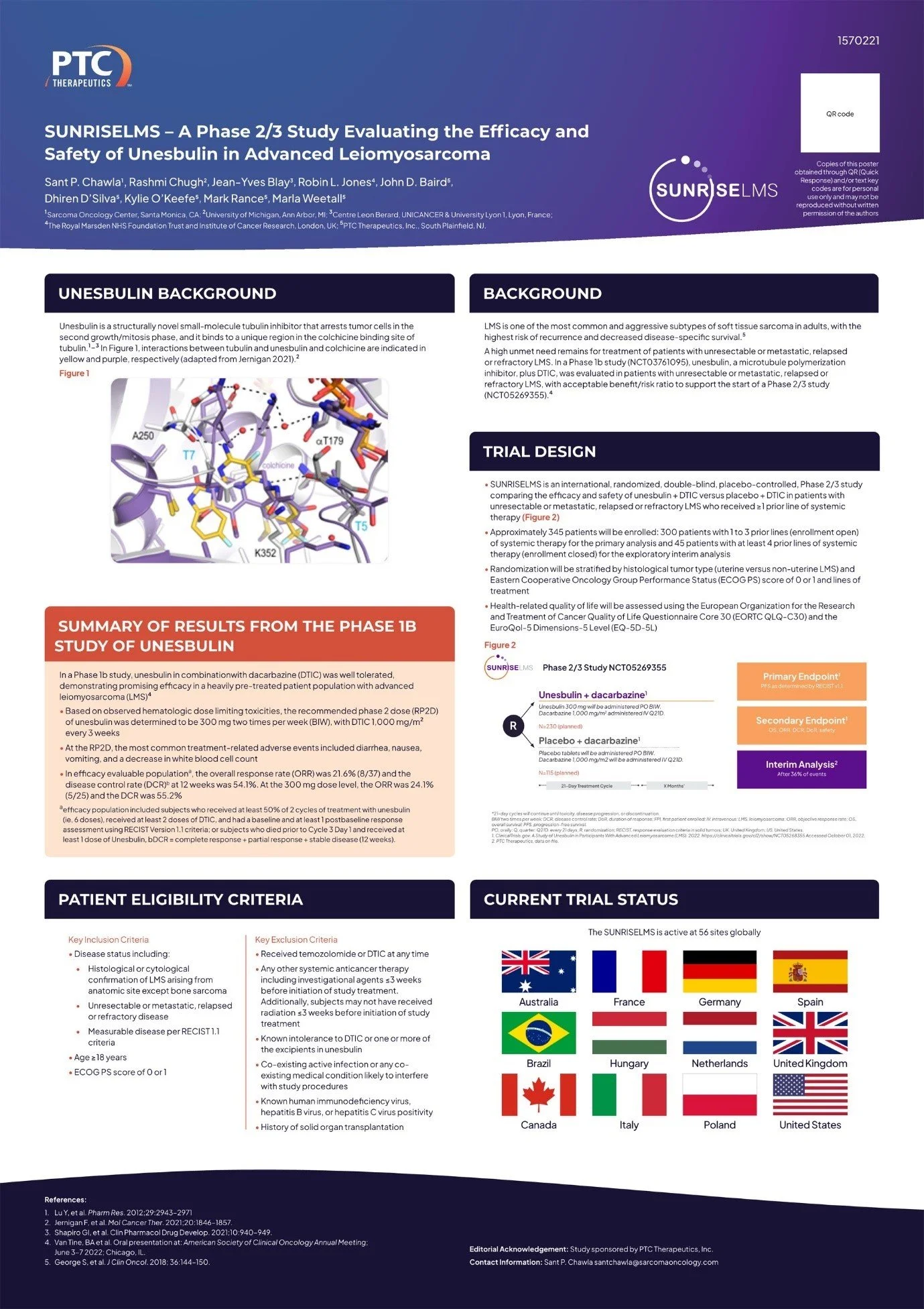
(P 548) A PHASE 2/3 STUDY EVALUATING THE EFFICACY AND SAFETY OF UNESBULIN IN ADVANCED LEIOMYOSARCOMA (SUNRISELMS)
Objective: Leiomyosarcoma (LMS) is one of the most common and aggressive adult soft tissue sarcoma subtypes, with a high risk of recurrence and decreased disease-specific survival (George S, et al. J Clin Oncol. 2018; 36:144–150). A high unmet need remains for treatment of patients with unresectable or metastatic, relapsed or refractory LMS. In a Phase 1b study (NCT03761095), unesbulin, a microtubule polymerization inhibitor, plus dacarbazine (DTIC), was evaluated in patients with unresectable or metastatic, relapsed or refractory LMS, with acceptable benefit/risk ratio to support the start of a Phase 2/3 study. Here, we report the study design of the Phase 2/3 study SUNRISELMS that will evaluate the efficacy and safety of the combination of unesbulin with DTIC in patients with advanced LMS who have received at least one prior line of systemic therapy (NCT05269355).
Methods: SUNRISELMS is an international, randomized, double-blind, placebo-controlled, Phase 2/3 study comparing the efficacy and safety of unesbulin + DTIC versus placebo + DTIC in patients with unresectable or metastatic, relapsed or refractory LMS who received ≥ 1 prior line of systemic therapy. Patients will be randomized 2:1 to unesbulin 300 mg orally twice weekly (BIW) in each 3-week treatment cycle plus DTIC 1000 mg/m2 intravenous (IV) once every 21 days (Q21D). Matching oral placebo BIW plus DTIC IV 1000 mg/m2 will be administered Q21D. An interim analysis will be performed when ~36%, or 88 of the progression-free survival (PFS) events occur.
Eligible patients will have unresectable or metastatic, relapsed or refractory, measurable LMS according to RECIST 1.1, and be ≥18 years. Approximately 345 patients will be enrolled: 300 patients with 1 to 3 prior lines of systemic therapy for the primary analysis and 45 patients with at least 4 prior lines of systemic therapy for the exploratory interim analysis. Randomization will be stratified by histological tumor type (uterine versus nonuterine LMS), ECOG score of 0 or 1 and lines of treatment.
The primary endpoint is PFS. Key secondary endpoints include overall survival, objective response rate, disease control rate, duration of response, and safety. The study is open and recruiting patients (NCT05269355).
Results: This section does not apply to this abstract since it is a trial in progress.
Conclusion: This section does not apply to this abstract since it is a trial in progress.